Life After Joint Replacement- What Recovery Really Looks Like
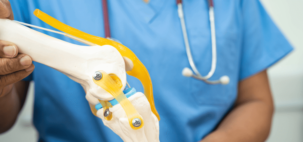
Life After Joint Replacement: What Recovery Really Looks Like? Joint replacement surgery is often seen as a life-changing step toward pain relief and improved mobility. However, many patients are unsure about what happens after the surgery. Recovery is not just about healing, it is a structured journey that involves rehabilitation, lifestyle adjustments, and gradual improvement. Understanding what to expect during joint replacement recovery Hisar helps patients stay prepared, motivated and confident throughout the process. The First Few Days After Surgery The initial phase of recovery begins immediately after surgery. Patients are usually encouraged to start gentle movement within a day to prevent stiffness and improve blood circulation. While there may be some discomfort, pain management techniques and medications help keep it under control. During this stage, medical teams focus on monitoring healing, preventing complications and helping patients regain basic mobility. Simple activities like sitting, standing, and taking a few steps are gradually introduced. This early phase is crucial because it sets the foundation for successful joint replacement recovery Hisar and prepares the body for further rehabilitation. The Role of Physiotherapy in Joint Replacement Recovery Hisar Physiotherapy is one of the most important components of recovery. It helps restore strength, flexibility and joint movement while ensuring proper healing. Structured physiotherapy after joint replacement Hisar programs begin soon after surgery and continue for several weeks or months. These programs include exercises to strengthen muscles around the joint, improve balance and enhance mobility. Consistency is key. Skipping sessions or not following prescribed exercises can slow progress and affect long-term results. With proper physiotherapy, patients can regain independence and return to daily activities more quickly. Post Knee Replacement Care and Daily Adjustments For patients undergoing knee surgery, following proper post knee replacement care Hisar guidelines is essential for smooth recovery. This includes maintaining proper posture, avoiding excessive strain and following movement precautions recommended by doctors. Daily routines may need temporary adjustments. Using support while walking, avoiding high-impact activities, and ensuring a safe home environment can help prevent falls or injuries. Patients are also advised to maintain a healthy diet and stay hydrated to support healing. Small changes in lifestyle during this phase contribute significantly to faster and safer recovery. Recovery Timeline: What to Expect Post Knee Replacement Care Hisar? Recovery after joint replacement varies from person to person, depending on age, overall health and commitment to rehabilitation. Most patients begin walking with support within a few days and gradually progress to independent movement. In the first few weeks, mobility improves steadily with regular physiotherapy. By 6 to 8 weeks, many patients can perform routine activities comfortably. Full recovery, including strength and flexibility, may take a few months. Understanding this timeline helps patients stay patient and focused during joint replacement recovery Hisar, avoiding unrealistic expectations and unnecessary stress. Returning to Normal Life and Activities One of the biggest goals of joint replacement surgery is to return to an active lifestyle. As recovery progresses, patients can gradually resume daily activities such as walking, driving and light exercise. However, it is important to avoid high-impact activities that may strain the joint. Low-impact exercises such as walking, swimming or cycling are recommended to maintain joint health. Following proper physiotherapy after joint replacement Hisar and medical advice ensures long-term success and prevents complications. With time and consistency, patients often experience improved mobility, reduced pain and better quality of life. Comprehensive Recovery Support at Orthomed Successful recovery depends not only on surgery but also on post-operative care and rehabilitation. For patients seeking reliable joint replacement recovery Hisar, Orthomed provides comprehensive support at every stage of the journey. From advanced surgical procedures to structured physiotherapy after joint replacement Hisar, Orthomed ensures that patients receive personalized care tailored to their needs. The hospital also offers guidance on post knee replacement care Hisar, helping patients recover safely and effectively. With experienced specialists, modern facilities, and a patient-focused approach, Orthomed supports faster healing, improved mobility, and long-term joint health. Their goal is to help patients return to an active, pain-free lifestyle with confidence. Final Thoughts Life after joint replacement is a gradual but rewarding journey. While the initial days may require patience and effort, consistent rehabilitation and proper care lead to significant improvements. Understanding the recovery process, following medical guidance and staying committed to physiotherapy are key to success. With expert care and structured support available at Orthomed, patients can confidently navigate joint replacement recovery Hisar and enjoy a healthier, more active life. Popular Post Understanding the Link Between Menopause and Bone Health in Women The Role of Robotics in Modern Orthopedic Surgery Plastic Surgery Post-operative care for neurosurgery patients: A Comprehensive Guide Surgical & Non-Surgical Trеatmеnts for Bonе Fracturе
Hand & Wrist Pain- Is It Fatigue or Something Serious
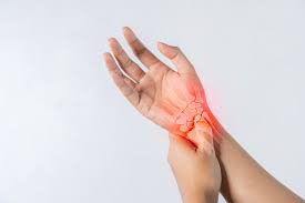
Hand & Wrist Pain: Is It Fatigue or Something Serious? Hand and wrist pain is often ignored as a minor issue caused by overuse or fatigue. In today’s digital world, where people spend long hours typing, scrolling, or performing repetitive tasks, discomfort in the hands and wrists has become increasingly common. While some cases are temporary and improve with rest, others may indicate underlying conditions that require medical attention. Understanding the difference between simple fatigue and a serious issue is essential. Knowing when to seek hand and wrist pain treatment Hisar can help prevent long-term complications and ensure proper recovery. Common Causes of Hand and Wrist Pain Hand and wrist pain can result from a variety of everyday activities. Repetitive movements such as typing, writing, lifting, or using tools can strain muscles and tendons over time. Poor ergonomics and incorrect posture also contribute to discomfort. In many cases, the pain is due to temporary fatigue and resolves with rest. However, persistent pain may be linked to conditions such as tendonitis, carpal tunnel syndrome, arthritis, or ligament injuries. If the pain continues despite rest or worsens over time, consulting an orthopaedic hand doctor Hisar is important to identify the exact cause and begin appropriate treatment. Signs That It’s More Than Just Fatigue & You Need Hand and Wrist Pain Treatment Hisar It can be difficult to determine whether pain is temporary or serious. However, certain symptoms indicate that medical attention is needed. Persistent pain lasting more than a few days, swelling, stiffness, numbness, or tingling in the fingers are warning signs. Weak grip strength, difficulty holding objects, or a feeling of instability in the wrist may also suggest underlying issues. Ignoring these symptoms can lead to worsening conditions and reduced functionality. Early evaluation for hand and wrist pain treatment Hisar helps prevent long-term damage and ensures faster recovery. Conditions That Affect the Hand and Wrist Several medical conditions can cause chronic hand and wrist pain. Carpal tunnel syndrome is one of the most common, caused by pressure on the median nerve, leading to numbness and tingling. Tendonitis occurs when tendons become inflamed due to overuse, causing pain and restricted movement. Arthritis can also affect joints in the hands and wrists, leading to stiffness and swelling. Ligament injuries or fractures from falls or accidents may cause severe pain and require immediate care. A qualified wrist pain specialist Hisar can diagnose these conditions accurately and recommend the right treatment plan. When to Visit a Wrist Pain Specialist in Hisar? Many people delay seeking medical care, assuming the pain will resolve on its own. However, early consultation can make a significant difference in treatment outcomes. You should visit a wrist pain specialist Hisar, if pain interferes with daily activities, affects sleep or does not improve with rest. Individuals experiencing recurring pain or symptoms like numbness and weakness should not ignore these signs. A specialist can perform a detailed examination and recommend suitable hand and wrist pain treatment Hisar options, including physiotherapy, medication, or advanced procedures if required. Treatment Options and Recovery Treatment depends on the severity and cause of the condition. Mild cases can often be managed with rest, splinting, ice therapy, and physiotherapy. Lifestyle changes & ergonomic improvements also play a key role in recovery. For more severe conditions, medical interventions such as injections or minor surgical procedures may be required. Early diagnosis ensures that treatment remains less invasive and more effective. Following a proper treatment plan and allowing adequate time for healing are essential to prevent recurrence and restore full functionality. Expert Hand and Wrist Care at Orthomed by Orthopedic Hand Doctor Hisar For individuals seeking reliable hand and wrist pain treatment Hisar, Orthomed offers advanced orthopedic care with a focus on accurate diagnosis and effective recovery. With experienced specialists, including a skilled orthopedic hand doctor Hisar, the hospital provides comprehensive treatment for conditions ranging from minor strains to complex wrist disorders. Patients benefit from personalized care plans designed to address the root cause of pain. As a trusted center with access to a qualified wrist pain specialist Hisar, Orthomed ensures modern treatment techniques, structured rehabilitation, and patient-focused care. Their goal is to help patients regain strength, mobility, and comfort in their daily activities. Final Thoughts Hand and wrist pain may seem like a minor inconvenience, but ignoring persistent symptoms can lead to serious complications. Knowing whether the pain is due to fatigue or an underlying condition is key to timely treatment. If discomfort continues or worsens, seeking expert care for hand and wrist pain treatment Hisar is the right step. With advanced solutions and experienced specialists at Orthomed, patients can achieve faster recovery and return to their normal routines without pain. Popular Post Understanding the Link Between Menopause and Bone Health in Women The Role of Robotics in Modern Orthopedic Surgery Plastic Surgery Post-operative care for neurosurgery patients: A Comprehensive Guide Surgical & Non-Surgical Trеatmеnts for Bonе Fracturе
Pediatric Orthopaedics: Signs Parents Should Never Ignore
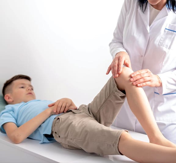
Pediatric Orthopaedics: Signs Parents Should Never Ignore Children are always on the move, running, playing, and exploring their environment. Though a certain number of bumps and bruises are part of growing up, there are some bone and joint problems that should not be overlooked. Early diagnosis of orthopedic issues is essential as the bones of children are still in the process of development, and undiagnosed disorders may have an impact on their future growth and locomotion. Knowledge of the time to consult an expert in the field of pediatric orthopaedics Hisar will assist parents to make sure that their child is growing strong, healthy, and active. Understanding Pediatric Orthopaedics and Why It Matters Pediatric orthopaedics is a specialty of treating and diagnosing bone, joint, and muscle diseases in children. Children, as opposed to adults, have growth plates, which are soft regions on the ends of the bones that allow their growth. These regions are more susceptible to damage and demand special treatment. Since the bones of children are still in development, the slightest problem can affect their future lives unless effectively addressed. This is the reason a visit to a children bone doctor Hisar is necessary in case the symptoms continue or even get worse. Early intervention not only helps to avoid complications but also to have proper alignment of the bones as well as development. Most of the pediatric orthopedic disorders can be cured with the aid of proper treatment, thus the children can resume their normal lives without having any lasting consequences. Persistent Joint Pain and Swelling in Children Children usually feel some discomfort following exercise, but continuous pain or swelling is worrisome. When a child keeps crying that he or she has pain in his or her knees, ankles, or any other joints, then it might show that there is a problem. crying that he or she has pain in his or her knees, ankles, or any other joints, then it might show that there is a problem. Ongoing discomfort can be provoked by conditions like infections, inflammation, or early joint disorders. In this situation, in-time treatment of child’s joint pain Hisar will count in preventing complications. Swelling, redness, or warmth around the joints should also be monitored by the parents. The signs can be signs of inflammation or harm that needs treatment. The denial of these symptoms may result in decreased mobility and chronic problems. Difficulty Walking or Limping One of the most obvious indicators that something can go amiss is limping. Although minor injuries may lead to temporary limping, lasting or unexplained limping can never be overlooked. A child who refuses to put weight on one leg or walks on one leg lopsided may have a fracture, muscle injury, or joint problem. At times, children might not be able to indicate the pain, and thus limping is the only observable symptom. Referring to a practitioner of paediatric orthopaedics, Hisar aids in diagnosing the cause beforehand. Such ailments like hip disorders, infections, or growth plate injuries are ailments which are easily treated when dealt with on time. Frequent Fractures or Bone Injuries Children are active by nature, and frequent fractures of the body or injuries might mean that the bones are weak or there are currently some underlying conditions. One fracture caused by a fall can be considered a normal case. However, multiple fractures after minimal impacts need to be assessed. Children bone doctor Hisar can check bone strength and diagnose nutritional deficiency or any medical condition that might cause problems with bone strength. Effective diagnosis of children can guarantee the right treatment of children, whether it is dietary counselling, supplements, or health care. Bone health should be taken care of at an early age to prevent any injuries in the future and enable the development of a robust skeleton. Delayed Growth or Physical Development Children develop differently; however, any significant physical developmental delays can be associated with orthopedic issues. The signs that are not to be overlooked include uneven length of limbs, inability to stand, late walking, or abnormal posture. These problems can be associated with bone issues, muscle weakness, or developmental problems. Early assessment in pediatric orthopaedics Hisar enables physicians to check the progress and prescribe remedies as necessary. Early intervention means that children grow their way to normal without having to face complications that could be treated in a more complicated manner in the future. Expert Child Joint Pain Treatment Hisar at Orthomed In the search for reliable care, Orthoped provides a variety of services specializing in pediatric orthopaedics Hisar, which offers extensive assessment and treatment of children with bone and joint diseases. Having qualified specialists and child-receptive care methods, Orthomed makes sure that young patients feel at home during treatment. It can be fractured care, child joint pain treatment Hisar, or mobility problems. The hospital offers individual solutions that are based on the needs of individual children. Being a trusted facility with access to a talented children bone physician Hisar, Orthomed is oriented to early diagnosis, effective treatment, and long-term health. They have a multidisciplinary approach to guarantee that children are taken care of, both during the first visit and recovery and follow-up. Final Thoughts The orthopedic health of children is crucial in their growth and development of the children. Although minor injuries are widely observed, there are specific symptoms that should not be overlooked, including constant pain, limping, frequent fractures, or slow growth. Timely attention to specialists of the field of pediatric orthopaedics Hisar guarantees the fact that the problems will be identified as soon as possible and effectively treated. In the presence of skilled assistance and developed care at the Orthomed, parents will be sure that their children are heading to a healthy and active lifestyle. Popular Post Understanding the Link Between Menopause and Bone Health in Women The Role of Robotics in Modern Orthopedic Surgery Plastic Surgery Post-operative care for neurosurgery patients: A Comprehensive Guide Surgical & Non-Surgical Trеatmеnts for Bonе Fracturе
From ACL Tear to Full Recovery: A Complete Guide for Athletes
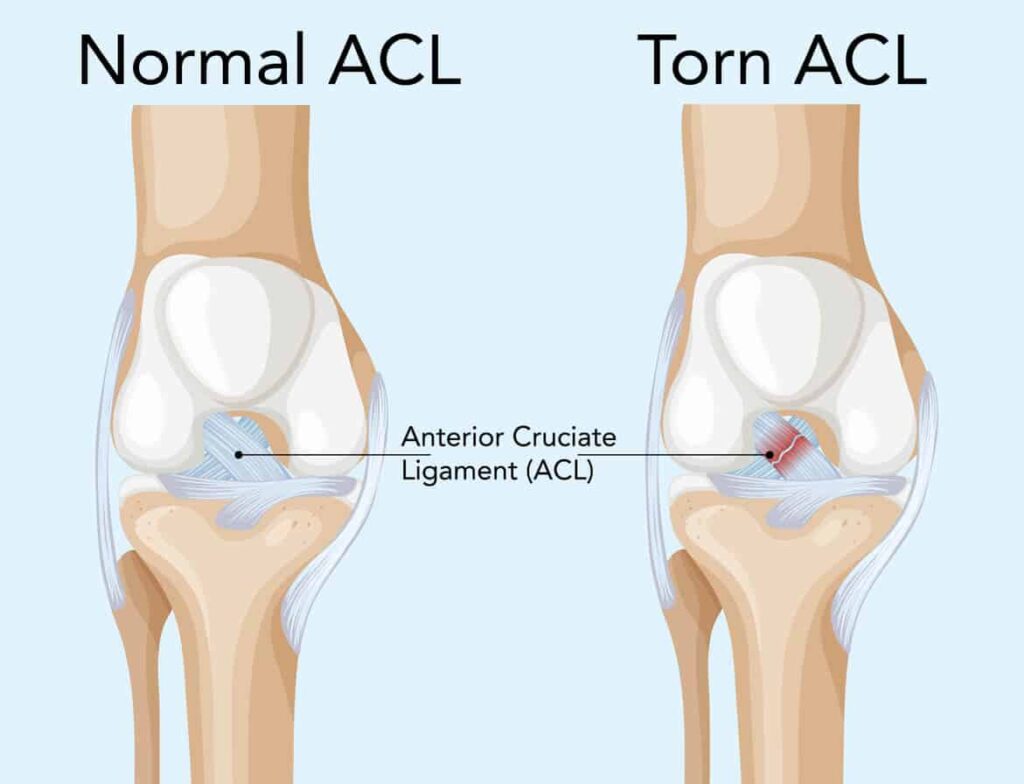
From ACL Tear to Full Recovery: A Complete Guide for Athletes One of the most severe and common injuries among athletes is an ACL (anterior cruciate ligament) tear. ACL injury can be a major performance and mobility hindrance, whether it happens in a sudden twist, pivot, or impact. But, through appropriate treatment and rehabilitation, the athletes can reappear. The knowledge of the path between injury and recovery is a must for any person who wants to achieve successful ACL tear treatment Hisar and safe recovery to sport. What Are ACL Injuries and Their Impact? ACL is a major ligament that provides stability to the knee joint, particularly in activities that involve sudden changes of direction, jumping, or high-impact activities. Laceration of this ligament causes instability, pain, and restricted knee activity. Athletes with an ACL tear complain of a popping sound on the injury and swelling with weight-bearing pain. It is worthwhile to have a specialist in the very near future to evaluate the severity level and start proper care. This early diagnosis is very important in deciding whether a non-surgical treatment or ligament injury surgery Hisar is necessary. Diagnosis and Treatment Options Physical examination and imaging tests like MRI scans are used in accurate diagnosis. These are useful in identifying the partial tear or complete rupture of the ligament. In minor injuries, simple treatment, such as rest, physiotherapy, and strengthening exercises, might be useful. Nevertheless, in the case of athletes or those who have complete tears, surgical reconstruction is usually advised. ACL tear treatment Hisar usually consists of reconstructing the damaged ligament with a graft and a planned rehabilitation. Early therapy guarantees improved stability and minimizes the risk of chronic problems. The Role of Ligament Injury Surgery Hisar in ACL Recovery Surgery is usually the choice for active individuals who desire to resume sports. Current ligament injury surgery Hisar procedures employ less invasive arthroscopic surgeries, which have small incisions and quicker recovery. Surgery aims to stabilize the knee and enable the patient to resume normal functioning. The development of surgical technology has led to accuracy, minimization of complications, and improved long-term outcomes. Surgery, however, is not sufficient; post-operative rehabilitation is key to successful recovery. Rehabilitation: The Key to Full Recovery with Knee Injury Recovery Hisar One of the most significant steps of ACL recovery is rehabilitation. It targets to build strength, knee flexibility, and balance. The healing process is a slow one and is normally broken into stages. The first phase is aimed at eliminating swelling and restoring limited movements. This is succeeded by strengthening exercises and later sport-specific training. Proper knee injury recovery Hisar programs help restore stability and functionality in the knee. Rehabilitation may be skipped or rushed, which can put people at risk of re-injury, and it is required that rehabilitation be consistent to be successful in the long term. Returning to Sports Safely The major worry of athletes after an ACL injury is how to resume the sport. It is natural to desire a fast comeback, but that may cause significant losses if it is premature. Safe return is reliant on several factors, such as muscle strength, joint stability, and fitness. Only when a doctor has given athletes the green light, and they have gone through a complete knee injury recovery Hisar program should they resume sports. Slow increase, training, and exercises for further strengthening are the measures that decrease the risk of future injuries. Expert ACL Care at Orthomed As an athlete who needs a good ACL tear repair Hisar, Orthomed is the place that provides a high-quality orthopedic service with attention to accuracy and healing. The hospital offers full service, including proper diagnosis, to highly qualified ligament injury surgery Hisar and rehabilitation. Orthomed has qualified experts and advanced surgical procedures that can guarantee successful care based on the individual needs of the patients. Their rehabilitation programs are designed to support complete knee injury recovery Hisar, helping athletes regain strength, confidence, and performance. The patient-centric approach of Orthomed will provide some form of guidance to individuals at each stage, including injury management and safe return to sports, which will make it a reliable option when it comes to ACL care in Hisar. Final Thoughts ACL tear may be a daunting blow, yet with proper ACL tear treatment Hisar, one can get back on track. Early diagnosis, appropriate treatment, successful surgery & regular rehabilitation are all essential in the healing process. It is better to prevent the usual pitfalls and adhere to the recommendations of the specialists to have a safe and successful recovery process. Advanced care and support at Orthomed will enable athletes to overcome injury, regain strength, and go back to their sport even better. Popular Post Understanding the Link Between Menopause and Bone Health in Women The Role of Robotics in Modern Orthopedic Surgery Plastic Surgery Post-operative care for neurosurgery patients: A Comprehensive Guide Surgical & Non-Surgical Trеatmеnts for Bonе Fracturе
Sports Injuries: Common Mistakes That Delay Recovery
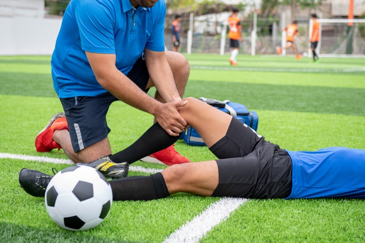
Sports Injuries: Common Mistakes That Delay Recovery Injuries in sports are prevalent among sportsmen and women in fitness and even among those who do physical activity more often. It can be a strain of the muscle, joint, or tear of a ligament; nevertheless, the recovery must be provided well and promptly. Nevertheless, numerous individuals make unknowingly wrong choices that decrease the rate of healing and predispose them to developing chronic problems. Such typical mistakes and wanting to find suitable sports injury treatment Hisar may assist in making sure that the recovery may be considered quicker and may be undertaken without recurrent problems. Ignoring Early Symptoms and Continuing Activity Among the most severe errors the individuals commit after getting an injury is disregarding the initial symptoms and resuming work. Slight pains, swelling, or pain are often ignored as being temporary, and people tend to persevere with the pain. But what would otherwise be a slight strain will soon become a severe injury unless it is meted out correctly. The absence of proper rest may increase tissue damage and increase the length of the recovery time by continuing the physical activity. A visit to a sports injury doctor Hisar before long will be sure to consult the injury that a doctor diagnoses and before it gets out of hand. Delaying Proper Diagnosis and Sports Injury Treatment Hisar A great number of people use homemade remedies or self-treatment without the help of an expert. Although simple treatment can assist during simple cases, severe cases like damage to ligaments should be handled by professionals. The side effects of not seeking medical attention include chronic pain, joint instability, and decreased mobility. Hisar case conditions, such as a ligament tear treatment Hisar are to be intervened promptly to guarantee appropriate recovery. Proper diagnosis with the help of imaging and specialist examination enables the physician to develop a specific treatment plan, which will save them time and further damage. Not Following a Structured Rehabilitation Plan This is not the end of the recovery when the pain has disappeared. Among the most prevalent ones is neglecting or failing to undergo physiotherapy and rehabilitation. Rehabilitation plays an important role in restoring strength, flexibility, and joint stability. In its absence, the affected region will be feeble, making it more likely to re-injure. Proper sports injury treatment Hisar comprises a systematic recovery program that progressively helps patients resume their regular level of activity safely and efficiently. Returning to Sports Too Soon Impatience during recovery is another major issue. When people begin to feel better, many people will resume sports or vigorous exercise without the body getting a chance to recuperate. It may cause re-injury that is usually more serious and takes even longer to recover. Muscles, ligaments, or joints that do not heal completely predispose them to incurring damage in the long term. Sports injury doctor Hisar, before going back to the activity, is a sure way of knowing that the body is prepared to withstand physical strain and minimise setbacks. Neglecting Proper Nutrition and Rest Just being treated by a doctor is not enough in curing disease. People also need to be fed well and get enough sleep. Most individuals do not appreciate the importance of nutrition and rest after an injury. A nutritious diet containing protein, vitamins, and minerals aids in repairing the tissues and decreasing inflammation. Hydration and sufficient sleep also play a vital role in recovery. The disregard of the mentioned factors can slow down the healing process and decrease the effectiveness of the best sports injury treatment Hisar plans. Expert Sports Injury Care at Orthomed with The Best Sports Injury Doctor Hisar In the case of people who have to solve problems related to sports injuries, the selection of medical assistance is crucial. Orthomed is a reliable facility that provides advanced sports injury treatment Hisar and enables patients to heal safely and efficiently. Orthomed offers high-quality care for injuries, taking into account both minor strains and more complex wounds of the ligaments, with experienced specialists and modern diagnostic and care facilities. And if ligament tear treatment Hisar or a visit to a professional sports injury doctor Hisar, the patient will be precisely diagnosed and treated individually at the hospital. The treatment strategy of Orthomed involves collaboration of medical skills, physiotherapy, and rehabilitation protocols in ensuring total recovery. Patients are guided through all levels of injury management to a safe return to sport, and long-term health and performance are ensured. Final Thoughts Sports injuries may be annoying, yet the appropriate strategy will mean a lot in the speed of recovery and results. It is necessary to avoid such common pitfalls as neglecting the symptoms, putting off treatment, overlooking rehabilitation, and resuming activity too soon. Early treatment and adherence to a developed course of action will guarantee a quicker recovery, and it will decrease the chances of complications. Patients may rely on recovery, and they can be assured of coming back to a stronger person with their active lifestyle, which they had before the expert support and advanced care, which is available at Orthomed. Popular Post Understanding the Link Between Menopause and Bone Health in Women The Role of Robotics in Modern Orthopedic Surgery Plastic Surgery Post-operative care for neurosurgery patients: A Comprehensive Guide Surgical & Non-Surgical Trеatmеnts for Bonе Fracturе
Back Pain vs Spine Disorder: When to Visit a Spine Specialist?
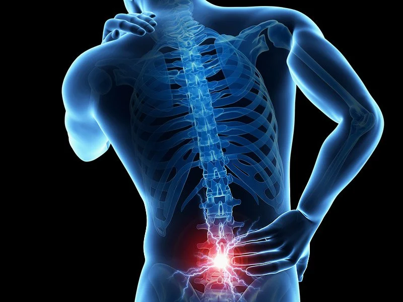
Back Pain vs Spine Disorder: When to Visit a Spine Specialist? One of the most widespread health complaints nowadays is back pain, which occurs in people of every age. Starting with long hours of sitting, there are posture issues, and physically demanding activities, among others, which are causing discomfort in the back. However, not all back pain is the same. Although rest may be enough to remove a temporary existence, other situations are indicative of an underlying spinal condition and need medical intervention. Knowing how to distinguish between normal back pain and a severe spine condition can help you know when to visit a spine specialist Hisar, and receive an appropriate treatment. Understanding Back Pain vs Spine Disorders From The View Of The Best Spine Specialist Hisar Strains on the muscles, poor posture, or small injuries are the most common causes of back pain. It tends to heal with rest, physiotherapy, or simple treatment. But when the pain is caused by structural issues within the spine, like disc issues, nerve compression, or degeneration of the spine, it is considered a spine disorder. Spinal disorders can be a herniated disc, spinal stenosis, spondylosis, or a problem with the nerves. Such cases need specific assessment and care from a spine specialist Hisar, and not general pain management. Early identification of this distinction can help avoid complications in the long-term and provide adequate care. Common Causes of Back Pain Back pain is a common occurrence in most people as a result of lifestyle factors. Some of the most common causes include poor posture, sitting for a long time, heavy lifting, and inactivity. In most instances, this kind of pain is temporary, and it subsides with rest, stretching, and simple back pain treatment Hisar options like physiotherapy or medication. Nevertheless, when the pain recurs or increases with time, it might be a symptom of a more serious spinal problem that needs additional research. Warning Signs of a Spine Disorder? Not all back pain is harmless. Certain symptoms suggest that the problem may be more serious and requires consultation with a spine specialist Hisar. Persistent pain lasting more than a few weeks, numbness or tingling in the arms or legs, weakness in muscles, difficulty walking or maintaining balance, and pain that radiates down the legs are key warning signs. Other red flags include pain that worsens at night, loss of bladder or bowel control, or severe stiffness limiting movement. These symptoms may indicate nerve compression or structural damage in the spine. In such cases, delaying treatment can lead to long-term complications, making early diagnosis essential. When Should You Visit a Spine Specialist in Hisar? Most of the patients do not know when to transition between general and specialized treatment. When resting, taking medicine, or physiotherapy is no longer helping your pain, then it is time to visit a spine specialist Hisar. The back pain should also be expertly consulted in case it disrupts daily life, sleep, or mobility. Symptoms should not be overlooked in individuals who have a history of injury, degeneration as a result of aging, or frequent episodes of pain. A specialist is able to conduct thorough assessments, such as imaging tests, to determine the underlying cause and prescribe the most efficient back pain treatment Hisar based on your condition. Treatment Options for Back Pain and Spine Disorders The cure is determined by the condition and its severity. In mild cases, non-surgical therapies like physiotherapy, posture repositioning, medications, and lifestyle modification may be effective. In more complicated spine disorders, more intricate treatment can be necessary. These may involve less invasive surgeries, injections, or, in worst-case scenarios, surgery. Modern back pain treatment Hisar aims at alleviating pain, regaining mobility, and preventing relapse. Early intervention ensures better outcomes and reduces the need for invasive procedures. Best Spine Hospital Hisar: Advanced Care at Orthomed When it comes to spine-related problems, it is important to select the appropriate healthcare facility. To those who are interested in expert treatment, Orthomed is considered one of the best spine hospital Hisar where the patients are provided with the best diagnosis and treatment against a broad spectrum of spinal disorders. Orthomed offers end-to-end care, including consultation and rehabilitation, with professional specialists and the latest technologies. The patients get to enjoy the benefits of accurate diagnosis, tailor-made treatment plans, and access to advanced procedures to recover. Be it simple back pain management Hisar or advanced spine care, Orthomed provides you with patient-centered care that focuses on restoring mobility and enhancing the quality of life. No matter if you need basic back pain treatment Hisar or specialized spine care, Orthomed ensures a patient-focused approach aimed at restoring mobility and improving quality of life. Final Thoughts Back pain is a small problem that may not be a big problem initially. However, neglecting the recurring symptoms may cause severe complications. The distinction between back pain in general and a spine disorder is important in order to receive timely treatment. When your pain persists or progresses or has an impact on your day-to-day activities, it is time to see a spine specialist Hisar. Orthomed has highly qualified specialists and modern facilities, which means that with proper treatment and professional attention, patients will be able to get healthy and resume a normal, active life. Popular Post Understanding the Link Between Menopause and Bone Health in Women The Role of Robotics in Modern Orthopedic Surgery Plastic Surgery Post-operative care for neurosurgery patients: A Comprehensive Guide Surgical & Non-Surgical Trеatmеnts for Bonе Fracturе
Robotic Joint Replacement: Technology Making Surgeries Safer
Robotic Joint Replacement: Technology Making Surgeries Safer Joint replacement has gone through a lot of changes, as it was initially a manual process, but at present, the processes are technologically advanced. Robotic-assisted surgery is one of the most significant innovations in orthopaedics that is changing the way joint replacements are conducted. Robotic knee replacement Hisar is becoming the number one choice among patients, with better precision, less risk, and quicker recovery. The present blog discusses the advantages of robotic technology in the field of surgery and its increasing popularity in Hisar. What Is Robotic Knee Replacement in Hisar? Robotic knee replacement Hisar is a new procedure in surgery that employs advanced robotic machines to help orthopedic surgeons be more precise in their joint replacement surgeries. Not as most people believe, the surgery is not done by a robot. The surgeon is in complete control during the surgery. The robotic system develops a detailed 3D image of the knee of the patient’s knee, which has the ability for accurate pre-surgical planning. It is used during surgery to guide the surgeon to make precise cuts and to place the implant in the right place based on the anatomy of the patient. Such a level of customization guarantees a higher level of alignment, enhanced long-term success, and better joint functioning. How Advanced Joint Surgery Hisar Improves Precision? Unparalleled precision is one of the benefits of advanced joint surgery Hisar with robotic assistance. Manual tools of alignment that are used in traditional techniques might not necessarily consider individual anatomic differences. Robotic systems, however, offer real-time information & instructions, making sure that the implant is inserted with the highest precision. This will minimize the likelihood of misalignment, which is a major cause of implant wear and failure in the long run. Precision also implies a reduced amount of damage to other tissues, leading to pain and faster recovery. This has led to increased patients choosing robot knee replacement Hisar instead of traditional methods. When Non-Surgical Treatments Stop Working? Patients have one of the greatest safety concerns when considering surgery. Surgery Robotic-assisted surgeries are a great way to increase the safety of surgery by reducing human errors and enhancing consistency. Using sophisticated joint surgery Hisar, surgeons are able to strategize all the steps to be undertaken during the operation before they go into the operating room. The robotic system is also capable of giving real-time feedback so that it can be adjusted during the surgery to achieve the best results. Less blood loss, less tissue damage, and smaller incisions are among the factors that lead to fewer complications. Moreover, accurate placement of the implants minimizes future revision surgeries. These aspects render robotic knee replacement Hisar a less risky and more secure choice among patients. Faster Recovery and Better Outcomes Recovery is a significant consideration that can affect a patient’s decision to undergo surgery. Robotic surgeries are meant to enhance quicker recovery and better mobility. Due to the increased accuracy and fewer invasions, patients have fewer side effects of the surgery, such as pain and swelling after surgery. This enables them to start physiotherapy at an earlier stage and recover faster. The patients who are subjected to robotic knee replacement Hisar frequently report greater stability of the knee joint, increased range of motion, and more natural motion than the traditional procedures. Robotic surgery is a great option among active individuals with structured rehabilitation and proper care. Most patients are back to normal routines in a few weeks. Who Should Opt For Knee Replacement Hisar? Robotic-assisted joint replacement can be applied to very diverse patients, particularly those with severe arthritis, joint deformities, or chronic knee pain affecting their daily life. It is especially helpful in cases when a person wants to have a specific, individual therapy and accelerate recovery. This approach is preferred by patients who desire long-term effects and better performance of the joint. To decide whether the advanced surgery of the joint using robotic technology should be performed or not, a consultation with an orthopedic specialist is possible, which will depend on your condition and lifestyle requirements. Orthomed Robotic Surgery Hisar: Advanced Care You Can Trust Robot knee replacement, Hisar is no exception, and the selection of the appropriate hospital is essential to get the most successful outcome. Orthomed robotic surgery Hisar is one of the leaders in the sphere of modern orthopedic treatment and surgery, providing the most innovative robotic-assisted technologies with emphasis on high accuracy and safety of patients. Orthomed uses the combined skills and expertise of surgeons with the latest robotic technology to provide highly precise and customized joint replacements. Patients are taken care of in all stages, including meticulous planning before surgery and well-organized rehabilitation programs. Being a reliable centre of high-quality joint surgery Hisar, Orthomed guarantees fewer complications, quicker recovery, and extended success. Their person-centered care can assist people who have lost their mobility to walk and engage in an active way of life once more with confidence, without any pain. Lastly Robotic-assisted joint replacement is transforming orthopedic treatment as it has made the surgeries safer, more accurate, and efficient. Being more accurate, quicker to recover, and better in the long run, it is fast becoming a more popular choice among patients. In case you are experiencing chronic knee pain and are considering surgery, you may want to look into robotic knee replacement Hisar, as it may be the first step to a pain-free life. Through professional services and the use of cutting-edge technology at Orthomed, patients have no reason to doubt their ability to take a safer and smarter road towards recovery. Popular Post Understanding the Link Between Menopause and Bone Health in Women The Role of Robotics in Modern Orthopedic Surgery Plastic Surgery Post-operative care for neurosurgery patients: A Comprehensive Guide Surgical & Non-Surgical Trеatmеnts for Bonе Fracturе
Joint Replacement Surgery: When Is It the Right Time?
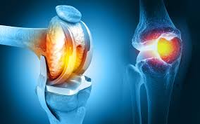
Joint Replacement Surgery: When Is It the Right Time? Joint pain is not just physical discomfort. It slowly impacts your independence, confidence, and overall quality of life. Be it the inability to move up the stairs easily, stand for long periods, or even get a good sleep, chronic joint problems tend to disrupt normal routines. Although most individuals attempt to cope with the symptoms over several years, they reach a point where they cannot cope with the symptoms using conservative treatments. Knowledge on when it is appropriate to think about joint replacement surgery Hisar can make you take an initiative step towards long-term relief and mobility. This guide will assist you in recognizing the important signs, knowing the treatment options, and making a wise choice. What Is a Joint Replacement Surgery? Joint replacement surgery refers to a medical operation whereby a part of the joint that is damaged or worn is substituted with artificial implants. These implants are designed to mimic the natural movement of the joint, decrease friction and pain due to bone-on-bone contact. Knee replacement Hisar and hip replacement surgeries are the most common procedures, and both are very effective in the treatment of advanced arthritis and degeneration of the joints. Advanced surgical methods at hip replacement hospital Hisar are aimed at accuracy, reduced tissue loss, and recovery rates, which make joint replacement safer and more readily available than ever. As the orthopedic care has advanced, patients now have a chance to receive a longer life of the implants, enhanced joint functionality, and faster recovery to normal life. Key Signs That Indicate You May Need Surgery at a Hip Replacement Hospital Hisar The reason why many patients postpone surgery is that they think that joint pains are normal aging. Nevertheless, some symptoms can give a clear indication that it is time to think of surgical intervention. One of the most frequent ones is persistent pain that has not gone away despite the medications or therapy. When your joint stiffness has limited movement, you find it difficult to walk, stand, and even when you are resting, you may still feel pain. Your joint status may have advanced a lot. Other red flags are decreased flexibility, swelling, joint instability, and the inability to perform the usual activities such as sitting, bending, or climbing stairs. These problems start to influence your way of life and autonomy, so it is necessary to visit a specialist to discuss joint replacement surgery in Hisar. When Non-Surgical Treatments Stop Working? Doctors usually recommend non-surgical options like physiotherapy, medicines, injections, and lifestyle modification before deciding on surgery. Such methods can give some short-term alleviation and assist in coping with initial joint issues. But these treatments can be ineffective with further loss of joint damage. The fact that they would delay recovery as the condition progresses may only be due to their continued use. Surgery is no longer a choice at this point, but the best method of replenishing joint function. Patients who promptly switch to surgical treatment usually have improved results than those who wait too long without the need to intervene. The Risks of Delaying Joint Replacement Surgery Hisar Postponing joint replacement may cause several complications that complicate treatment. The more the joint is damaged, the weaker the muscles surrounding it are, which are not used. This may impact balance, posture, and general physical strength. There can also be deformities due to severe joint deterioration, which can complicate surgery and prolong the recovery period. Altered body mechanics can result in the development of other problems in other joints or back pain in the patient in some cases. Early intervention not only stops such complications but also enables patients to heal more quickly and become better able to move around. Who Should Consider Knee Replacement Hisar? No longer are elderly patients confined to joint replacement. Although age may be a consideration, much of this depends on how badly the joints are damaged, how much pain, and how much lifestyle is impaired. Ideal candidates include people who have advanced arthritis, sports injuries, or degenerative joint conditions that disrupt normal life. With the durability of implants and surgical methods, young patients are increasingly choosing procedures such as knee replacement Hisar. The comprehensive analysis of an orthopedic specialist can assist in making a decision about the need for surgery and the type of intervention that will have the most favorable outcome. Advanced Joint Replacement Care at Orthomed The selection of the appropriate hospital is an important step towards success. Orthomed provides the best orthopedic solutions in a well-equipped and trained team that is ready to offer patients the best joint replacement surgery Hisar. The Orthomed is a reputable knee and hip surgery hospital Hisar because it has developed expertise in both knee and hip surgeries. The hospital has high-quality care, proper diagnosis, surgical planning, and programmed rehabilitation. Being highly accurate, patient safety, and quicker recovery, Orthomed makes sure that each patient is given a unique form of treatment that is aimed at regaining mobility and quality of life. The patient-based approach that is provided by the hospital assists individuals in getting back to active living. We provide initial consultation to post-surgery support. Final Thoughts Making such a big decision to have a joint replacement surgery is life-changing when one makes the right move at the right time. The presence of persistent pain, lack of progress or improvement with non-surgical interventions is a clear indicator that surgery could be a requirement. Patients can prevent complications and obtain a more positive outcome by making a timely intervention and seeking the assistance of qualified specialists. With more than just basic care services in Orthomed, the next step towards joint replacement surgery Hisar can be the move towards a pain-free, active, and happier life. Popular Post Understanding the Link Between Menopause and Bone Health in Women The Role of Robotics in Modern Orthopedic Surgery Plastic Surgery Post-operative care for neurosurgery patients: A Comprehensive Guide Surgical & Non-Surgical Trеatmеnts for Bonе Fracturе
Rheumatoid Arthritis vs Osteoarthritis: What Patients in Hisar Need to Know
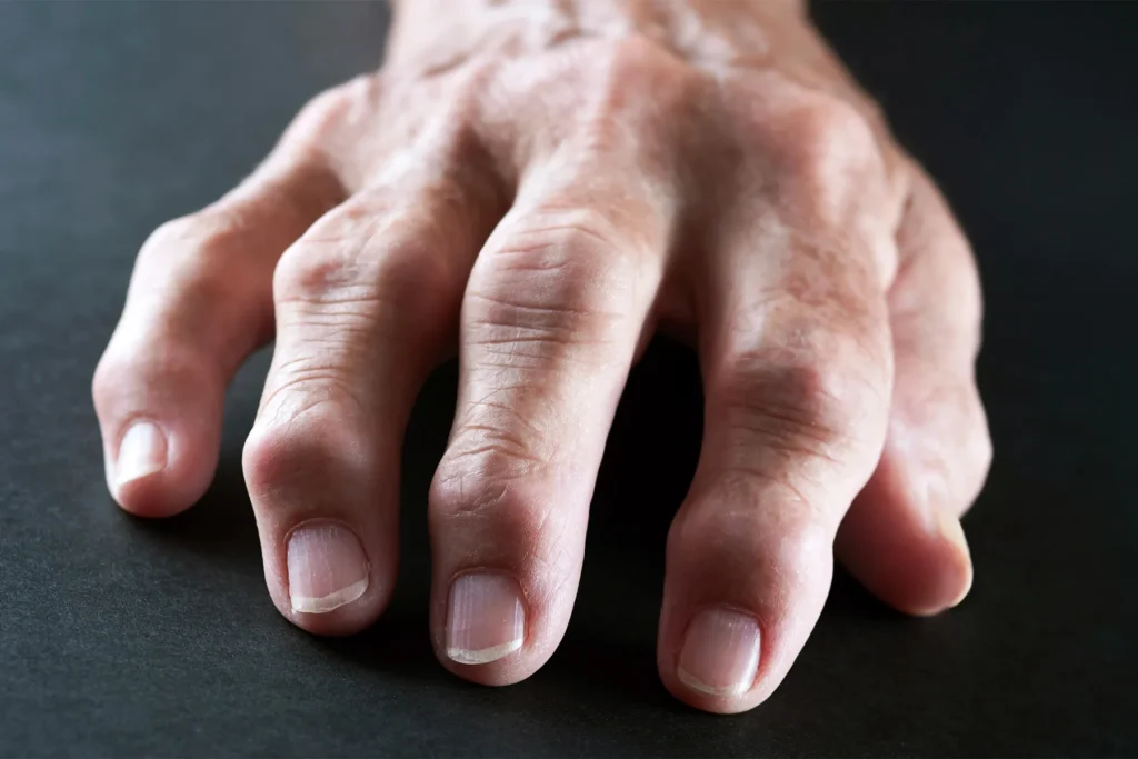
Rheumatoid Arthritis vs Osteoarthritis: What Patients in Hisar Need to Know Joint pain is one of the most common health concerns affecting people of all ages. However, not all arthritis is the same. Two of the most frequently diagnosed types are rheumatoid arthritis and osteoarthritis. While both conditions cause joint pain and stiffness, their causes, progression, and treatment approaches are very different. For patients seeking clarity and effective care, understanding the difference arthritis types Haryana is essential. With rising awareness and access to specialized care, many individuals are now consulting an arthritis specialist Hisar to get an accurate diagnosis and personalized treatment. What is Arthritis? More Than Just Joint Pain Arthritis is a broad term used to describe inflammation of the joints. It can affect mobility, cause pain, and significantly impact daily activities. The two most common forms, rheumatoid arthritis and osteoarthritis, differ in how they develop and affect the body. Osteoarthritis is primarily a degenerative condition caused by wear and tear of joint cartilage over time. Rheumatoid arthritis, on the other hand, is an autoimmune disorder where the body’s immune system mistakenly attacks its own joints. Recognizing these differences is the first step toward effective treatment. What Is Rheumatoid Arthritis? Rheumatoid arthritis Hisar patients often experience a condition where the immune system attacks the lining of joints, causing inflammation, swelling, and pain. It is a chronic autoimmune disease that can affect multiple joints simultaneously. Unlike osteoarthritis, rheumatoid arthritis can affect people at a younger age and may involve other organs such as the skin, eyes, and lungs. Common symptoms include persistent joint swelling, morning stiffness lasting more than an hour, fatigue, fever, and symmetrical joint pain (affecting both sides of the body). Early diagnosis and autoimmune joint pain treatment Hisar Haryana are crucial to prevent joint damage and disability. What Is Osteoarthritis? Osteoarthritis is the most common form of arthritis and is often associated with aging. It occurs when the protective cartilage cushioning the ends of bones gradually wears down, leading to bone-on-bone contact. This condition typically affects weight-bearing joints such as the knees, hips, and spine. Unlike rheumatoid arthritis, osteoarthritis usually develops slowly over time. Symptoms include joint pain during movement, stiffness after inactivity, reduced flexibility, and a grinding sensation in the joints. Key Differences Between Rheumatoid Arthritis and Osteoarthritis Understanding the difference arthritis types Haryana helps patients seek the right treatment. Rheumatoid arthritis is an autoimmune condition, while osteoarthritis is degenerative. Rheumatoid arthritis often affects multiple joints symmetrically, whereas osteoarthritis usually affects specific joints based on wear and tear. Morning stiffness in rheumatoid arthritis tends to last longer, while in osteoarthritis it typically improves within a short period. Rheumatoid arthritis may also involve fatigue and systemic symptoms, which are not common in osteoarthritis. Because the causes differ, treatment approaches also vary significantly. Causes and Risk Factors Rheumatoid arthritis is linked to immune system dysfunction, genetic factors, and environmental triggers. Women are more commonly affected than men. Osteoarthritis is primarily caused by aging, joint overuse, obesity, previous injuries, and lifestyle factors. Repetitive strain on joints increases the risk over time. Understanding risk factors helps patients take preventive steps and seek timely care from an arthritis specialist Hisar. Diagnosis: Why Accurate Evaluation Matters? Proper diagnosis is essential to differentiate between these two conditions. Doctors evaluate symptoms, perform physical examinations, and use diagnostic tests such as blood tests, X-rays, and MRI scans. Blood tests can detect markers of inflammation and autoimmune activity, which are important in diagnosing rheumatoid arthritis. Imaging tests help assess joint damage and cartilage loss in osteoarthritis. Consulting an experienced arthritis specialist Hisar ensures accurate diagnosis and appropriate treatment planning. Treatment Approaches for Rheumatoid Arthritis Treatment for rheumatoid arthritis focuses on controlling inflammation, preventing joint damage, and improving quality of life. Medications such as disease-modifying anti-rheumatic drugs (DMARDs), biologics, and anti-inflammatory drugs are commonly used. Physiotherapy and lifestyle changes also play an important role. Early autoimmune joint pain treatment Hisar Haryana can slow disease progression and prevent long-term complications. Treatment Approaches for Osteoarthritis Osteoarthritis treatment focuses on pain relief, improving joint function, and maintaining mobility. Lifestyle changes such as weight management, exercise, and physiotherapy are key components. Medications may be used to manage pain and inflammation. In advanced cases, surgical options such as joint replacement may be recommended to restore mobility and reduce pain. Living with Arthritis: Managing Daily Life Both rheumatoid arthritis and osteoarthritis require long-term management. Regular exercise, balanced nutrition, and maintaining a healthy weight help reduce joint stress. Patients should avoid prolonged inactivity, as it can worsen stiffness. Using supportive devices and following medical advice can improve comfort and mobility. Emotional well-being is equally important, as chronic pain can affect mental health. Support from family, healthcare providers, and therapy programs can help patients stay motivated. Expert Arthritis Care at Orthomed For patients in Hisar, Orthomed offers comprehensive care for both rheumatoid arthritis and osteoarthritis. With experienced specialists and advanced diagnostic facilities, the hospital provides personalized treatment plans tailored to each patient’s condition. Whether you need early diagnosis, ongoing management, or advanced treatment options, Orthomed ensures holistic care for joint health. Their approach combines medical expertise, physiotherapy, and lifestyle guidance to improve patient outcomes. As a trusted center for rheumatoid arthritis Hisar care and overall joint health management, Orthomed helps patients regain mobility and maintain a better quality of life. Prevention and Early Intervention While rheumatoid arthritis cannot always be prevented, early diagnosis and treatment can control symptoms and prevent joint damage. For osteoarthritis, maintaining a healthy lifestyle, avoiding joint overuse, and staying physically active can reduce risk. Early consultation with an arthritis specialist Hisar helps identify issues before they become severe. Final Thoughts Understanding the difference between rheumatoid arthritis and osteoarthritis is essential for effective treatment and long-term joint health. While both conditions cause pain and stiffness, their causes and management approaches are very different. If you are experiencing persistent joint discomfort, swelling, or reduced mobility, seeking timely medical advice is crucial. With access to expert care and advanced treatment options at
Cerebral Palsy Treatment in Hisar: Best Orthopaedic Care for Children at Orthomed

Cerebral Palsy Treatment in Hisar: Best Orthopaedic Care for Children at Orthomed Cerebral palsy (CP) is a group of neurological conditions that affect movement, posture, and muscle coordination in children. While it is a lifelong condition, early diagnosis and the right medical support can significantly improve mobility, independence, and quality of life. With growing awareness and advanced care options, families are now actively seeking cerebral palsy treatment Hisar that combines medical expertise with long-term rehabilitation. This guide helps parents understand CP, its symptoms, and how specialized orthopedic care can support their child’s development. What Is Cerebral Palsy? Cerebral palsy is caused by damage to the developing brain, usually before or during birth. This affects muscle control, coordination, and sometimes speech and cognition. The severity can vary widely, from mild movement difficulties to significant physical challenges. Although CP cannot be cured, early intervention and structured therapy can help children achieve better functional outcomes. Common Symptoms of Cerebral Palsy Symptoms may appear in infancy or early childhood. Some children may show delayed developmental milestones such as late sitting, crawling, or walking. Others may have stiff or floppy muscles, poor coordination, or difficulty maintaining balance. Additional signs include involuntary movements, abnormal posture, difficulty with fine motor skills, and speech challenges. If parents notice these symptoms, consulting a cerebral palsy doctor Hisar early is essential. Importance of Early Diagnosis and Intervention Early diagnosis plays a critical role in managing cerebral palsy effectively. The earlier therapy begins, the better the chances of improving mobility and preventing complications. A qualified CP specialist Haryana evaluates muscle tone, movement patterns, and developmental milestones to create a personalized treatment plan. Early intervention can reduce muscle stiffness, improve coordination, and support overall growth. Orthopaedic Challenges in Cerebral Palsy Children with CP often face orthopedic issues such as muscle tightness, joint deformities, contractures, and bone alignment problems. These can affect walking ability and posture. Orthopedic care focuses on correcting these physical challenges, improving movement, and preventing long-term complications. Timely intervention helps children become more independent in daily activities. Cerebral Palsy Treatment Options Treatment for CP is multidisciplinary and tailored to each child’s needs. It often includes physiotherapy, occupational therapy, and, in some cases, surgical intervention. Physiotherapy helps improve muscle strength, flexibility, and coordination. Occupational therapy focuses on daily skills such as dressing, writing, and self-care. Speech therapy may also be included when needed. Orthopedic procedures may be recommended to correct deformities, release tight muscles or improve joint alignment. These interventions are planned carefully by specialists to enhance mobility and comfort. Role of Orthopaedic Surgery in CP Care In certain cases, surgery plays an important role in improving function and quality of life. Procedures may include tendon lengthening, muscle release, or bone correction. These surgeries aim to reduce pain, improve posture, and enable better movement. Post-surgical rehabilitation is essential to achieve optimal results. Comprehensive CP Care at Orthomed For families seeking a trusted best CP care centre Hisar Haryana, Orthomed offers specialized orthopedic care for children with cerebral palsy. The hospital provides a multidisciplinary approach, combining orthopedic expertise with rehabilitation support. With experienced specialists and child-friendly care protocols, Orthomed focuses on improving mobility, correcting deformities, and enhancing independence. From diagnosis to long-term management, families receive continuous support tailored to their child’s needs. As a center for advanced cerebral palsy treatment Hisar, Orthomed ensures each child receives personalized care aimed at maximizing their potential. Long-Term Management and Support Cerebral palsy requires ongoing care and monitoring. Regular physiotherapy, assistive devices, and periodic medical evaluations help children maintain progress and adapt to growth changes. Family involvement is crucial. Parents and caregivers play a key role in supporting therapy routines and encouraging active participation in daily activities. Helping Children Lead Independent Lives With the right care and support, many children with CP can lead active and fulfilling lives. Early intervention, consistent therapy, and specialized orthopedic care help improve mobility and confidence. Encouraging participation in school, social activities, and physical exercises further supports emotional and mental well-being. Conclusion Cerebral palsy may present lifelong challenges, but with early diagnosis and expert care, children can achieve significant improvements in mobility and independence. Seeking timely consultation with a cerebral palsy doctor Hisar, and working with a qualified CP specialist Haryana ensures the best outcomes. With advanced care and dedicated support available at Orthomed, families in Hisar can access reliable and compassionate cerebral palsy treatment Hisar, helping their children grow stronger and live more confident, active lives. FAQs What is the main cause of cerebral palsy? Cerebral palsy is caused by damage to the developing brain, usually before or during birth, affecting movement, muscle control, and coordination. At what age can cerebral palsy be diagnosed? Signs may appear in infancy, and diagnosis is often made between 6 months and 2 years of age by a cerebral palsy doctor Hisar. Can cerebral palsy be cured? There is no cure, but early cerebral palsy treatment Hisar with therapy and medical care can significantly improve mobility and quality of life. What treatments help children with cerebral palsy? Treatment may include physiotherapy, occupational therapy, medications, and sometimes surgery under a CP specialist Haryana. How can I choose the best CP care centre in Hisar? Look for experienced specialists, multidisciplinary care, and rehabilitation support. A best CP care centre Hisar Haryana, like Orthomed, provides comprehensive treatment and long-term management. Popular Post Understanding the Link Between Menopause and Bone Health in Women The Role of Robotics in Modern Orthopedic Surgery Plastic Surgery Post-operative care for neurosurgery patients: A Comprehensive Guide Surgical & Non-Surgical Trеatmеnts for Bonе Fracturе